This is another video based on the procedures we reviewed during the OPPE session from May 6, 2024. This one is on the use of the transvenous pacer in the ED.
Placement of the CORDIS in the IJ
The first step is placing a Cordis line in the internal jugular under ultrasound guidance.
Once the vessel is located and punctured, follow the steps in the video below for placement of the Cordis catheter.

Setting up the pacer box and floating the pacer line
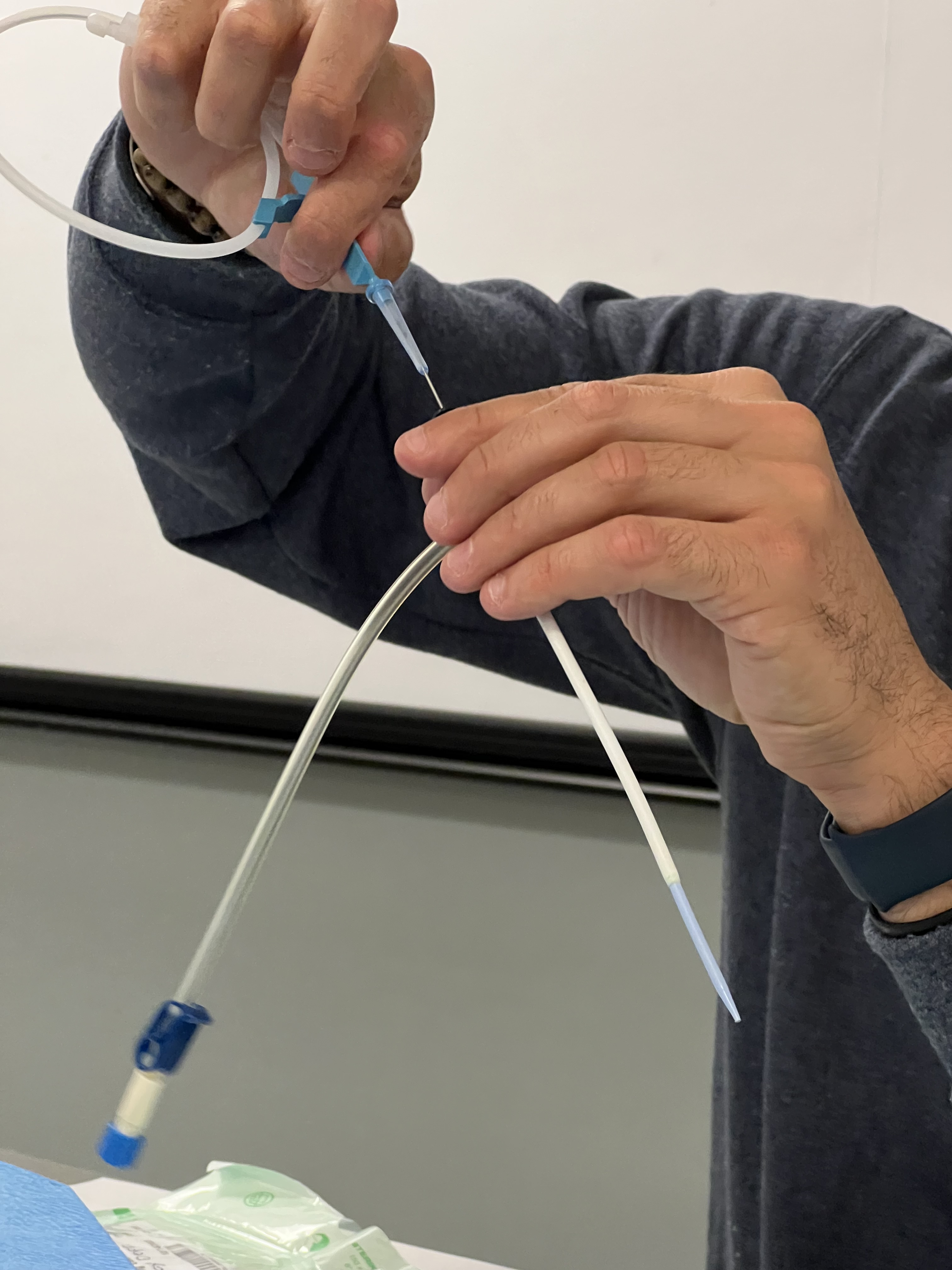
Once the Cordis is in and secured, it can be used to float the pacer line into the right ventricle. This can be done under ultrasound guidance (sub-xiphoid view by a helper) or watching for an injury pattern on the monitor. You’ll also need a non-sterile helper to adjust the pacer box.
There are a lot of steps in floating the wire and setting up the box.
- Get proper informed consent
- Use sterile drape and technique (preferably on the right IJ, or the left subclavian — though save that for the permanent pacer later)
- Place the cordis in the IJ (see above)
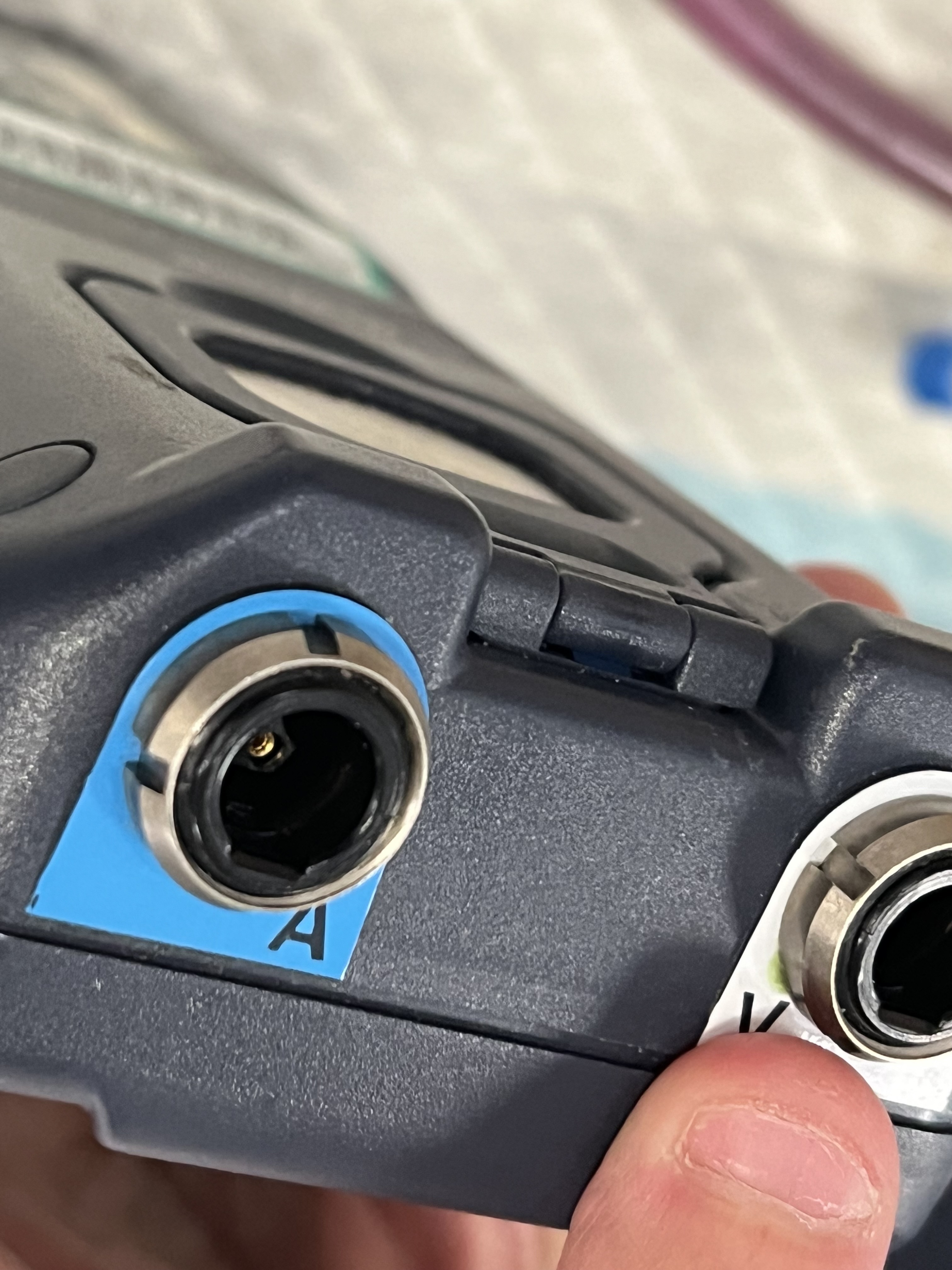
- Non-sterile helper will plug the wires into the ventricular port
- Feed the wire through the cords port (be sure to match the curve to how you want it to get into the RV)
- Once at the double-line (20 cm mark) connect the adapter pins to the end of the wire and hand to the non-sterile helper to connect to the pacer’s wire in the ventricular port.
- Now push the wire down to the triple-line (30 cm mark) then inflate the balloon (using the correct syringe).
- Now float the wire down further until you see it in the RV on US or injury pattern on the V1 lead of the cardiac monitor.
- Once in place, check the pulse-ox for pulse (or someone can palpate the pulse).
- Set the atrial output to the minimum needed for capture.
- Deflate the balloon & lock its stopcock.
- Move down the sterile sleeve and connect to the Cordis.
- Secure in place with sutures and a sterile dressing.


You must be logged in to post a comment.