This can be visualized in both a short and long view. Pain in the tendon, good story plus ultrasound can be diagnostic. Track the tendon down using the probe. Here’s the long view with an intact and ruptured tendon.
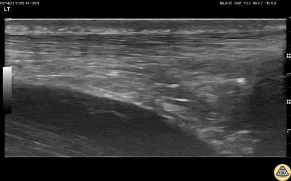

Here’s the short view.


Shoulder
Shoulder dislocations can also be visualized on ultrasound. Start posterior and visualize the humerus then move it upward and medially until you can see the humeral head.

 In this picture you can see the rotator cuff (red), humeral head (green) and glenoid (purple).
 And here is the shoulder rotating
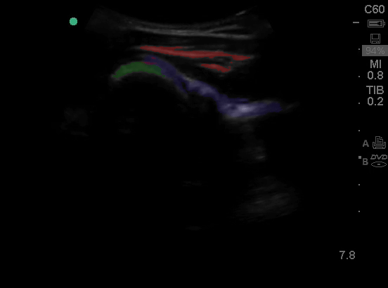


And what a dislocated shoulder looks like, space between the glenoid and the humeral head.


And if you want to block the shoulder, use the nerve block box which has everything in it except meds. There’s also a nerve block order set. Put the patient on telemetry. Use 10 cc lidocaine 1% and 10 cc of saline, inject with a block needle or a spinal needle. Inject into the space between the humeral head and glenoid.
- bupivicaine 10-20 cc
- decadron 4 mg
For peripheral nerve blocks you’ll need 3-5 mL. For pain blocks, like the fascia iliaca, you’ll need 20-30 mL. Don’t exceed the maximum dose and use the linear probe.

You must be logged in to post a comment.